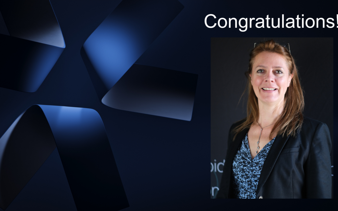
Publications
Association of Autofluorescence-Based Detection of the Parathyroid Glands During Total ThyroidectomyWith Postoperative Hypocalcemia Risk Results of the PARAFLUO Multicenter Randomized Clinical Trial
We evaluated the feasibility and the tolerance of repeated fluorescent indocyanine green angiography in free flap monitoring, and determined the intraoperative predictive values of flap vitality.
Reconstructive surgery has for objective to restore anatomical defects using flaps. We tested fluorescent angiography in reconstructive surgery using the Fluobeam™ device for flap planning and monitoring.
Indocyanine green fluorescence (ICGF) and parathyroid autofluorescence (AF) are two new techniques that aid in the identification of parathyroid glands (PG) intraoperatively during thyroidectomy. There is no study comparing the efficacy of these techniques.
Intraoperative localisation and preservation of parathyroid glands improves outcomes following thyroid and parathyroid surgery. This can be facilitated by fluorescent imaging and methylene blue; a fluorophore is thought to be taken up avidly by parathyroid glands.
Parathyroid gland (PG) identification during thyroid and parathyroid surgery is challenging. Accidental parathyroidectomy increases the rate of postoperative hypocalcaemia. Recently, autofluorescence with near infrared light (NIRL) has been described for PG visualization. The aim of this study is to analyze the increased rate of visualization of PGs with the use of NIRL compared to white light (WL).
Parathyroid glands (PGs) can be particularly hard to distinguish from surrounding tissue and thus can be damaged or removed during thyroidectomy. Postoperative hypoparathyroidism is the most common complication after thyroidectomy. Very recently, it has been found that the parathyroid tissue shows near-infrared (NIR) auto-fluorescence which could be used for intraoperative detection, without any use of contrast agents.
Identification of parathyroid glands may be challenging during thyroid and parathyroid surgery. Accidental resection of the glands may increase the morbidity of the surgery. The aim of this study was to evaluate accuracy in identification of autofluorescent parathyroid glands with the use of near infrared light in real time.
Lymphedema is a chronic peripheral swelling caused by a dysfunction of the lymphatic system, leading to discomfort and loss of upper limb movement. Therapies to treat or manage this swelling have limited evidence, partly because of a paucity in objective lymphatic measures. This study explored the role of near-infrared (NIR) imaging in evaluating interventions.
Vascularized lymph node transfer (VLNT) as a surgical treatment for cancer-related lymphedema has shown beneficial effects. The mechanism of action of this procedure involves lymphangiogenesis and lymphovenous communications (LVC) within the lymph nodes.
It is proposed that direct mechanical loading can impair dermal lymphatic function, contributing to the causal pathway of pressure ulcers. The present study aims to investigate the effects of loading on human dermal lymphatic vessels. Ten participants were recruited with ages ranging from 24 to 61 years.
Impaired lymph formation and clearance has previously been proposed as a contributory factor in the development of pressure ulcers. The present study has been designed to trial fluorescence lymphangiography for establishing how lymphatic function is altered under a clinically relevant form of mechanical loading.
The Fluobeam® is a portable, near-infrared camera that is held and controlled by the surgeon to visualize tissue perfusion using indocyanine green (ICG) fluorescence imaging.
The presence of midline sub-umbilical and/or suprapubic scar can sometimes hinder breast reconstruction using deep inferior epigastric perforator (DIEP) procedure.
The aim of the study was to compare 2 methods of the sentinel lymph node biopsy (SLNB) procedure in bladder cancer: we applied technetium radiocolloid (RadCol) detected by a gamma ray detection probe, and indocyanine green (ICG) detected by a near-infrared fluorescent (NIRF) camera.
Sentinel lymph node (SLN) detection techniques have the potential to change the standard of surgical care for patients with prostate cancer. We performed a lymphatic mapping study and determined the value of fluorescence SLN detection with indocyanine green (ICG) for the detection of lymph node metastases in intermediate- and high-risk patients undergoing radical prostatectomy and extended pelvic lymph node dissection.
Sentinel node biopsy (SNB) is an established method in oral squamous cell carcinoma (OSCC) for staging the cN0 neck and to select patients who will benefit from a neck dissection. Near-infrared fluorescence (NIRF) imaging has the potential to improve the SNB procedure by facilitating intraoperative visual identification of the sentinel lymph node (SN).
The aim of this study was to compare lymphatic drainage patterns detected with fluorescent dye indocyanine green (ICG) with the lymphatic drainage patterns detected with radiotracer (99m)Tc-nanocolloid in dynamic sentinel node biopsy (DSNB) procedures.
There is some controversy about the value of sentinel lymph node excision (SLNE) in patients with head and neck malignancies. The gold standard for detection and targeted extirpation of the SLN is lymphoscintigraphy with (99m)Tc-nanocolloid.
IMPORTANCE: The metastatic status of regional lymph nodes is the most relevant prognostic factor in breast cancer, melanoma, and other solid organ tumors with a lymphatic spread. The current gold standard for detection and targeted excision of the sentinel lymph node is preoperative lymphoscintigraphy with technetium Tc 99m.